Quick answer
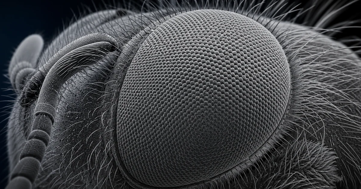
An SEM image of a bee eye reveals the compound eye as a tightly packed field of small optical units called ommatidia. Each unit contributes part of the bee's visual field, and together they create a system optimized for motion detection, navigation, flower recognition, and orientation during flight.
For an image atlas entry, the bee eye is one of the clearest examples of why scanning electron microscopy is powerful for biological surfaces. The structure is both visually striking and technically meaningful: the eye is not a smooth dome, but a patterned array of lenses, ridges, hair bases, and microscale surface features.
What the SEM image reveals
The main feature is the compound eye surface. In a well-prepared specimen, the SEM view shows hundreds or thousands of ommatidia as repeated polygonal facets. Many appear hexagonal because hexagonal packing is an efficient way to cover curved surfaces with minimal wasted space.
At higher magnification, the boundaries between ommatidia become clear. You may also see interfacetal hairs or bristle-like structures, depending on the species, the preparation, and the region of the eye. These hairs can influence airflow, particle capture, and light management. They also make the image more informative because they give a direct sense of scale and surface relief.
The surface texture matters. SEM can show whether the lenses are clean, collapsed, dusty, coated, cracked, or affected by preparation artifacts. A good bee eye SEM image is therefore both a biological image and a small lesson in sample handling.
Likely imaging mode
The most likely mode is secondary electron imaging. Secondary electrons are highly surface sensitive, so they are excellent for showing the shape of ommatidia, the depth of grooves, and the position of hairs. This is why most classic insect SEM images have a sculptural, high relief appearance.
Backscattered electron imaging is less common for this subject unless the goal is to highlight compositional contrast. For example, a metal coating, mineral contamination, or debris could appear with different brightness in a backscattered electron detector. For the eye structure itself, secondary electron imaging is usually the better first choice.
Low accelerating voltage can help preserve surface detail and reduce charging. A conductive coating, such as gold, gold-palladium, platinum, or carbon, is commonly used because insect cuticle is not naturally conductive enough for stable high-vacuum imaging.
Sample preparation considerations
Bee eyes contain curved cuticle, delicate hairs, and biological material that can deform during drying. Preparation usually starts with careful cleaning and fixation. If the goal is publication-quality morphology, the specimen may be dehydrated through an ethanol series and dried using critical point drying or another method that reduces collapse from surface tension.
Mounting angle matters. A straight top-down image emphasizes the repeating pattern of ommatidia. An oblique image reveals curvature, facet height, and hairs more dramatically. Coating thickness also matters. Too little coating may cause charging. Too much coating can soften the finest details and make individual lens boundaries less sharp.
Common artifacts include collapsed facets, dust, charging streaks, broken hairs, and coating grains. These artifacts do not make the image useless, but they should be recognized before drawing biological conclusions.
Why the structure matters
The bee eye is central to pollination biology. Bees use vision to detect flowers, navigate landscapes, recognize polarized light, and stabilize flight. SEM does not show how the photoreceptors work inside the eye, but it shows the external architecture that supports the optical system.
The repeated ommatidial pattern is also useful for teaching SEM image interpretation. It introduces surface topography, magnification, working distance, sample tilt, charging, and biological sample preparation in a single familiar organism.
Related SEM terms
- Compound eye
- Ommatidium
- Secondary electron imaging
- Interfacetal hair
- Conductive coating
- Critical point drying
- Biological SEM
- Surface topography