Subject
SEM Troubleshooting
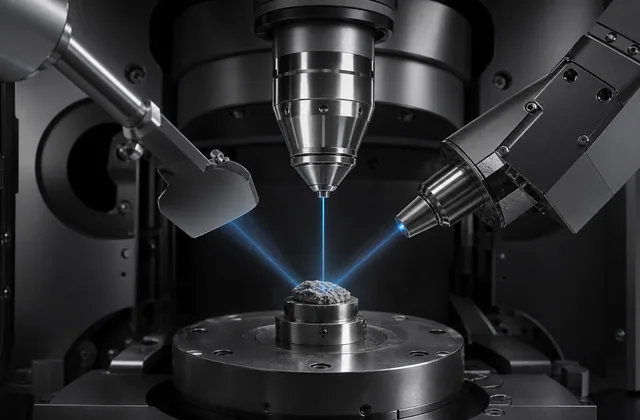
Future hub for charging, contamination, drift, astigmatism, poor signal, beam damage, and EDX artifacts.
Quick answer
To fix charging, first confirm that the contrast moves, blooms, streaks, or drifts with beam exposure. Then reduce dose, improve grounding, lower keV, use variable pressure if available, or coat the sample when compatible.
Key takeaways
- Charging is often solved by several small changes, not one magic setting.
- Lower keV helps many samples but can reduce signal or EDX usefulness.
- Do not mistake charging contrast for composition.
What it means
How to Fix Charging in SEM is best understood as a practical SEM decision point. It affects how the image is formed, how the sample behaves under the beam, and how confidently the resulting contrast can be interpreted.
The concept should always be tied to the sample and question. SEM settings that work beautifully on a conductive metal fracture surface may fail on a polymer, biological specimen, powder, or hydrated material.
Why it matters in SEM
SEM is powerful because it links surface detail, signal generation, detector geometry, and sometimes elemental analysis. That also means image quality can be misleading when settings are chosen by habit.
For how to fix charging in SEM, the operator should document the detector, accelerating voltage, working distance, vacuum mode, beam current or spot size, sample preparation, and any coating. Those details turn a picture into interpretable evidence.
Practical interpretation
Start with a low magnification survey, find representative areas, then move toward the feature of interest. Change one major condition at a time so the cause of improved or degraded contrast remains clear.
When the image changes, ask whether the change reflects the sample or the instrument setup. Charging, contamination, beam damage, detector shadowing, and preparation artifacts can all create convincing but false stories.
Common mistakes
- Treating magnification as the main measure of quality.
- Omitting detector and beam settings from notes.
- Using high beam energy when surface detail or beam sensitivity is the real issue.
- Interpreting brightness without considering detector type.
- Forgetting that preparation can create the structure being imaged.
Operator checklist
- Define the sample question before choosing settings.
- Capture survey and detail images.
- Record SEM metadata with every final image.
- Recheck focus and stigmation at final magnification.
- Compare at least two conditions when the interpretation is uncertain.
Where to go next
A short editorial reading list. Pick whichever fits how you like to learn.
- Royal Microscopical Society: professional microscopy community and education
- Microscopy Society of America: society resources, meetings, and microscopy community