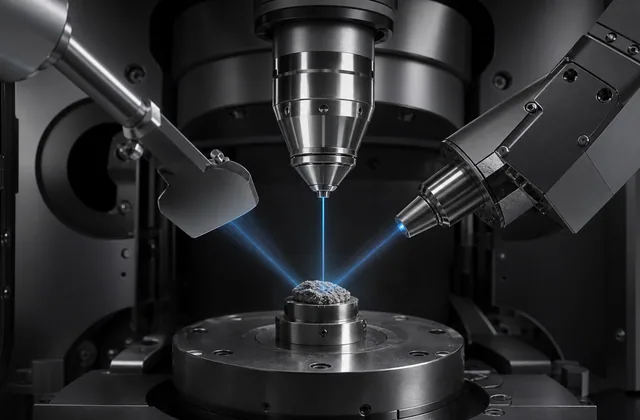
Subject
SEM Detectors and Signals
Technical resources on secondary electrons, backscattered electrons, in-lens detectors, SE1, SE2, and detector selection.
Quick answer
Secondary electron detectors and backscatter electron detectors answer different SEM questions.
Use secondary electron, or SE, imaging when you need surface morphology, texture, edges, and a visually intuitive topographic image. Use backscatter electron, or BSE, imaging when you need contrast related to composition, average atomic number, phases, inclusions, grains, or orientation effects.
Neither signal is universally superior. A strong SEM workflow often uses SE to find and understand the surface, then BSE to test whether brightness differences are related to material differences.
Signal origin
Secondary electrons are low-energy electrons emitted from near the specimen surface. They usually provide high surface sensitivity and strong topographic contrast.
Backscattered electrons are primary beam electrons that enter the specimen, undergo elastic scattering, and leave the specimen with relatively high energy. Because the probability of backscattering increases with atomic number, BSE imaging can reveal material contrast that is nearly invisible in SE mode.
What SE detectors show best
SE detectors are often the first choice for:
- surface morphology
- particles and fibers
- fracture surfaces
- insect and biological surfaces
- coatings and contamination
- microfabricated structures
- navigation at low and medium magnification
SE images can be beautiful and immediately readable. They often show relief, pores, ridges, cracks, and edges with strong apparent depth.
The caution is that brightness in an SE image is not simply height, composition, or conductivity. It is affected by detector position, local electric fields, sample tilt, charging, edge emission, and the mixture of SE1 and SE2 signal.
What BSE detectors show best
BSE detectors are often the first choice for:
- multiphase alloys
- polished geological sections
- ceramics and composites
- inclusions and precipitates
- heavy element contamination
- phase distribution before EDX
- orientation contrast in crystalline materials
In many BSE images, heavier regions appear brighter and lighter regions appear darker. For example, a heavy metal inclusion in a lighter matrix may stand out clearly.
BSE imaging is especially useful before EDX analysis because it helps locate compositionally distinct regions worth measuring.
Comparison table
| Feature | Secondary electron detector | Backscatter electron detector |
|---|---|---|
| Main contrast | Surface topography and edges | Composition, average atomic number, phase, orientation |
| Signal energy | Low | High relative to SE |
| Surface sensitivity | Very high | Lower than SE, depends on beam energy and sample |
| Spatial resolution | Often higher for surface detail | Often lower because signal comes from a larger interaction volume |
| Best specimen geometry | Rough or textured surfaces | Flat, polished, or moderately smooth samples |
| Common detector position | Chamber side, in-lens, through-the-lens | Annular or segmented detector near pole piece |
| Typical first use | Find morphology | Find phases or material contrast |
Detector placement
SE detectors are frequently mounted in the chamber or integrated into the electron column. A classic Everhart-Thornley detector collects secondary electrons using a biased grid and scintillator. In-lens and through-the-lens systems collect secondary electrons near or inside the column and are often favored for high-resolution imaging.
BSE detectors are commonly placed below the pole piece, often as annular or segmented solid-state detectors. Their position favors collection of high-energy electrons leaving the specimen at relatively high angles.
Segmented BSE detectors can also create directional topographic contrast. By comparing segments, the operator can distinguish some topographic effects from average atomic number contrast.
Practical selection guidance
If the sample is rough, unknown, or biological, start with SE. It will usually give the fastest visual understanding of the surface.
If the sample is polished, multiphase, geological, metallurgical, ceramic, or compositionally complex, start with BSE or capture BSE immediately after SE.
If brightness differences in SE imaging may be composition-related, verify with BSE and EDX. SE alone is usually not enough to make a compositional claim.
If brightness differences in BSE imaging may be caused by topography, reduce tilt, improve polishing, lower the working distance if appropriate, and compare detector segments or SE images.
BSE and atomic number contrast
The backscatter coefficient generally rises with atomic number. This is why BSE is widely used for Z-contrast imaging.
The useful phrase is "average atomic number contrast." A phase containing heavier elements tends to appear brighter than a phase dominated by lighter elements, assuming comparable geometry and imaging conditions.
There are limits. BSE brightness is not a complete chemical analysis. Density, crystallography, surface angle, beam energy, detector geometry, and charging can all influence the image. Use EDX or another analytical method when elemental identity matters.
SE and topographic contrast
SE images are often interpreted as morphology because low-energy secondary electrons escape from near the surface. Edges and features facing the detector commonly appear bright.
This visual style is useful, but it is also why SE images can exaggerate roughness. Two features with the same material and height can appear different if one faces the detector and the other is shadowed.
For quantitative height, use profilometry, AFM, stereo SEM methods, or calibrated 3D workflows rather than raw SE brightness.
When to use both
Many serious SEM sessions should include both SE and BSE images of the same field of view.
Use the paired images this way:
| Observation | Likely interpretation |
|---|---|
| Bright in SE, not distinct in BSE | Topography, edge effect, charging, or local geometry |
| Bright in BSE, not especially raised in SE | Higher average atomic number or phase contrast |
| Bright in both | Could be a raised feature, heavier material, or both |
| Dark in SE and BSE | Depression, light phase, poor collection geometry, or charging |
Notes on beam energy
Higher accelerating voltage increases interaction volume and often strengthens BSE production. This can improve compositional contrast but reduce surface specificity.
Lower accelerating voltage improves surface sensitivity and can reduce charging or beam damage in some samples. It may also reduce X-ray generation for EDX and change the BSE contrast mechanism.
There is no universal best voltage. Choose beam energy according to the signal you need, the sample thickness and conductivity, and whether imaging or analysis is the priority.
Bottom line
SE is the morphology workhorse. BSE is the composition and phase contrast workhorse.
For research-grade interpretation, use them as complementary signals. SE tells you what the surface looks like. BSE tells you whether the material itself may be changing.
Where to go next
A short editorial reading list. Pick whichever fits how you like to learn.
- Royal Microscopical Society: professional microscopy community and education
- Microscopy Society of America: society resources, meetings, and microscopy community