Quick answer
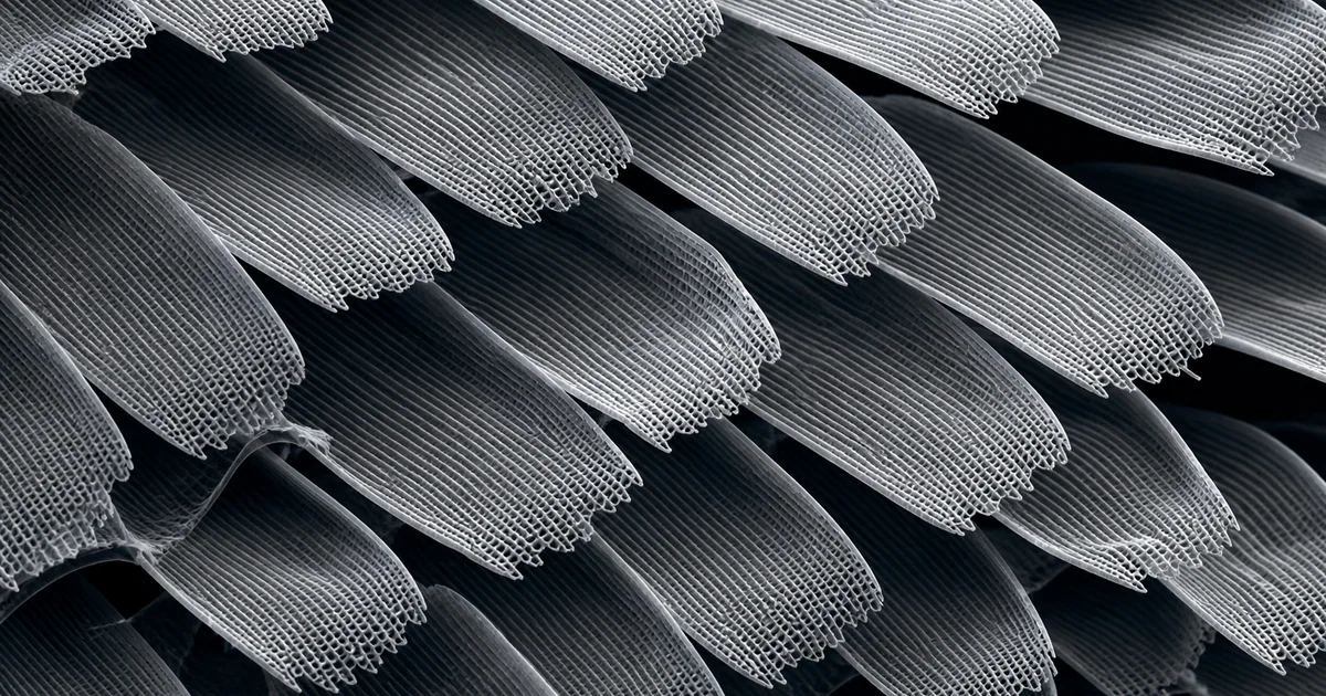
An SEM image of butterfly wing scales reveals a layered surface made of many overlapping microscopic scales. At higher magnification, each scale can show ridges, cross ribs, pores, windows, and fine lamellar structures. These details are central to how butterfly wings interact with light, water, heat, and air.
Butterfly wing scales are a classic SEM subject because they look delicate but highly engineered. The image is immediately recognizable, yet it also opens the door to advanced topics such as structural color, biomimetic surfaces, and nanoscale patterning.
What the SEM image reveals
At low magnification, the wing surface often looks like roof tiles or shingles. The scales overlap in organized rows, with their free edges pointing in a consistent direction. This arrangement protects the wing membrane and creates a textured surface.
At higher magnification, the individual scales become the main subject. SEM can show long parallel ridges, cross ribs connecting the ridges, small pores, and sometimes more complex internal lamellae. These structures can influence how light is scattered, reflected, or filtered. In some butterflies, nanoscale architecture contributes to vivid structural color, even though the SEM image itself is grayscale.
The image may also show damage. Missing scales, broken ribs, dust, adhesive residue, and handling marks are common. Because the structures are thin and lightweight, sample handling has a major effect on the final image.
Likely imaging mode
Secondary electron imaging is the most likely mode for butterfly wing scales. It provides strong surface topography and edge detail, which is exactly what is needed for ridges, ribs, and overlapping scale margins.
Backscattered electron imaging is less common for general scale morphology. It can be helpful if the study is looking for particles, mineral contamination, or coating-related contrast. EDX may be used in special cases, but it is not the usual first tool for understanding wing scale structure.
Low accelerating voltage is often preferred because wing scales are delicate, thin, and prone to charging. A thin conductive coating can stabilize imaging while keeping the microstructure visible.
Sample preparation considerations
The main preparation goal is to preserve the natural scale arrangement. Butterfly wings can often be mounted dry, but they should be handled gently because scales detach easily. A small piece of wing can be placed on carbon tape or a stub adhesive, with care taken to avoid pressing the surface flat.
Orientation changes the story the image tells. A broad top view shows scale overlap and row organization. A tilted view can reveal scale thickness and edge structure. A fractured or edge-on view may expose internal scale architecture, but it is harder to prepare and interpret.
Coating must be thin and uniform. Excessive coating can obscure pores and fine lamellae. Too little coating may cause charging, especially around lifted scale edges. Charging can create bright patches, streaks, or apparent texture that is not part of the biological surface.
Why the structure matters
Butterfly wing scales are more than decoration. Their microstructure can contribute to color, hydrophobicity, thermal regulation, camouflage, signaling, and flight surface behavior. SEM makes the physical basis of these functions visible.
The subject is also important in materials science. Researchers study butterfly scales as models for photonic structures, anti-wetting surfaces, lightweight materials, and microstructured coatings. A single SEM image can connect entomology, optics, and biomimetic engineering.
Related SEM terms
- Wing scale
- Structural color
- Secondary electron imaging
- Lamellar microstructure
- Cross rib
- Hydrophobic surface
- Charging artifact
- Biomimetic material