Quick answer
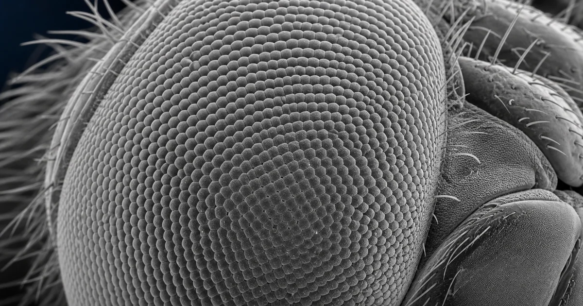
An SEM image of a dragonfly eye shows a large compound eye covered with dense arrays of ommatidia. Each ommatidium is a small optical unit, and the complete eye gives the dragonfly wide visual coverage that supports hunting, obstacle avoidance, and rapid flight control.
Dragonfly eyes are among the most recognizable insect structures under scanning electron microscopy. They are large enough to be easy to mount and dramatic enough to show why SEM is so useful for surface morphology. At the same time, they are technically rich because the eye surface changes curvature, facet density, and viewing angle across the specimen.
What the SEM image reveals
The dominant feature is the repeating lens array. The ommatidia often appear as tightly packed polygonal facets over a curved surface. In a strong SEM image, the viewer can see the boundaries between lenses, the smoothness or texture of individual facets, and the way the array follows the shape of the head.
A dragonfly eye may show regional variation. Some areas can have larger or differently organized facets than others, reflecting visual specialization. SEM does not directly measure vision, but it can reveal the external organization that supports optical performance.
At higher magnification, the image may show fine surface deposits, collapsed areas, scratches, or preparation marks. These details are useful because they separate real biological structure from artifact. A beautiful SEM image is not automatically a clean biological record. Interpretation still requires attention to prep and imaging conditions.
Likely imaging mode
Secondary electron imaging is the most likely mode. It gives strong contrast from surface topography, which is exactly what is needed to show the ommatidial field. The grooves between facets, the edges of the lenses, and the overall curvature are all made easier to read by secondary electron contrast.
Backscattered electron imaging has a smaller role unless there is a reason to look for compositional differences. For example, backscatter may highlight contamination, coating thickness differences, or foreign particles. For normal morphology, secondary electrons provide the more intuitive image.
Because dragonfly eye surfaces are curved, depth of field is a major advantage of SEM. A moderate working distance and suitable aperture can help keep more of the surface in focus while preserving enough detail for the facet pattern.
Sample preparation considerations
The eye should be cleaned gently and dried without collapsing the surface. Critical point drying is often preferred for delicate biological structures because it reduces distortion caused by liquid surface tension during drying. If the specimen is already dry, it may still need careful handling to avoid dust and mechanical abrasion.
Mounting angle is important. A low magnification overview can show the eye's scale and curvature. A tilted high magnification view can emphasize relief and facet boundaries. The best atlas images often include one broad view and one closer view, because the structure is meaningful at both scales.
Conductive coating is usually required. Insect cuticle can charge under the electron beam, especially at high vacuum. A thin metal coating improves stability, but excessive coating can blur fine boundaries and make the lens surface look grainy.
Why the structure matters
Dragonflies are visual predators. Their compound eyes support fast tracking of prey and environmental motion. SEM helps readers see the physical surface of this visual system, especially the dense packing and curvature that are hard to appreciate with unaided sight.
The dragonfly eye also gives researchers and students a clean bridge between biological form and instrument technique. It demonstrates surface imaging, depth of field, specimen tilt, coating, magnification, and the difference between visual pattern and analytical interpretation.
Related SEM terms
- Compound eye
- Ommatidium
- Secondary electron detector
- Working distance
- Depth of field
- Biological SEM
- Conductive coating
- Surface topography