Quick answer
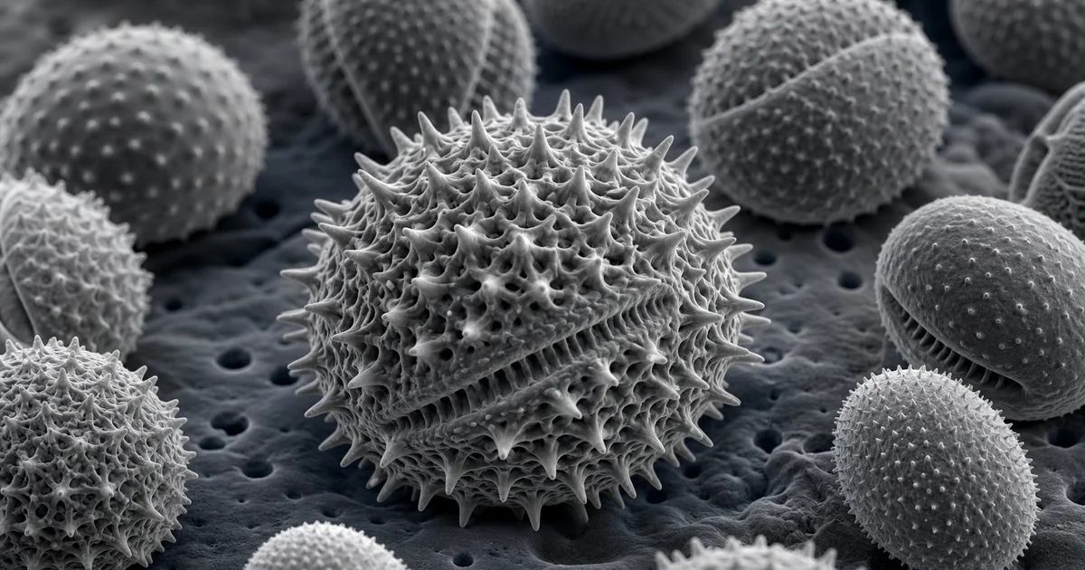
An SEM image of pollen shows the outer wall of pollen grains in remarkable detail. The most important visible features are the exine surface, apertures, pores, furrows, spines, ridges, and reticulate or net-like patterns. These structures can be highly diagnostic, which is why pollen is a classic subject for scanning electron microscopy.
Pollen is also an unusually good SEM teaching sample. It is small, often robust, visually varied, and rich in surface texture. A single image can explain magnification, depth of field, surface topography, biological identification, and specimen preparation.
What the SEM image reveals
The main structure is the pollen grain wall. The outer layer, called the exine, may be smooth, spiny, pitted, ridged, netted, folded, or covered with complex sculpturing. SEM is especially useful because it shows the three-dimensional surface of this wall rather than only a transmitted-light outline.
Many pollen grains also show apertures. These may appear as pores, furrows, or thinner regions in the wall. Apertures matter because they are involved in pollen germination and because their number and arrangement are important in pollen classification.
In a mixed sample, SEM can show how different pollen types vary in size, shape, and surface ornamentation. However, a striking image is not the same as a complete identification. Species-level interpretation requires comparison with reference collections and knowledge of where the sample came from.
Likely imaging mode
Secondary electron imaging is the standard mode for pollen morphology. The reason is simple: pollen analysis is mostly about surface form. Secondary electrons resolve ridges, pits, spines, pores, and subtle wall texture with strong topographic contrast.
Backscattered electron imaging may be useful when the question involves contamination, mineral dust, coating variation, or particles attached to the pollen surface. EDX can also be used if there is a need to check inorganic residue, although it is not normally required for basic pollen morphology.
Low accelerating voltage is often helpful because pollen grains are small and can charge. A thin conductive coating improves image stability while preserving fine surface texture.
Sample preparation considerations
Pollen can be easier to prepare than many soft biological tissues, but it still needs care. Dry pollen can often be mounted directly on carbon adhesive and coated for SEM. Wet or chemically treated samples may require dehydration before imaging.
The biggest practical problem is contamination. Dust, fibers, adhesive strings, and loose debris can look confusing at high magnification. A clean mount and gentle transfer are important. Pollen grains should be separated enough that individual shapes and apertures remain visible.
Coating thickness matters. A heavy metal coating improves conductivity but can fill small pits or make delicate exine features look rounded. Too little coating may lead to charging, which can create streaking, brightness shifts, or image drift.
Why the structure matters
Pollen morphology has value in botany, ecology, allergy research, archaeology, and forensic science. The surface pattern can help identify plant groups, reconstruct environments, track pollination, or link materials to a location.
SEM is especially valuable because many pollen features are surface features. Light microscopy can show size and general shape, but SEM can reveal the sculptural detail that makes the grain recognizable. For readers, pollen is a useful reminder that SEM is not only for metals, chips, and nanomaterials. It is also a major tool for biological surfaces.
Related SEM terms
- Pollen exine
- Aperture
- Palynology
- Secondary electron imaging
- Surface ornamentation
- Conductive coating
- Charging
- EDX contamination check