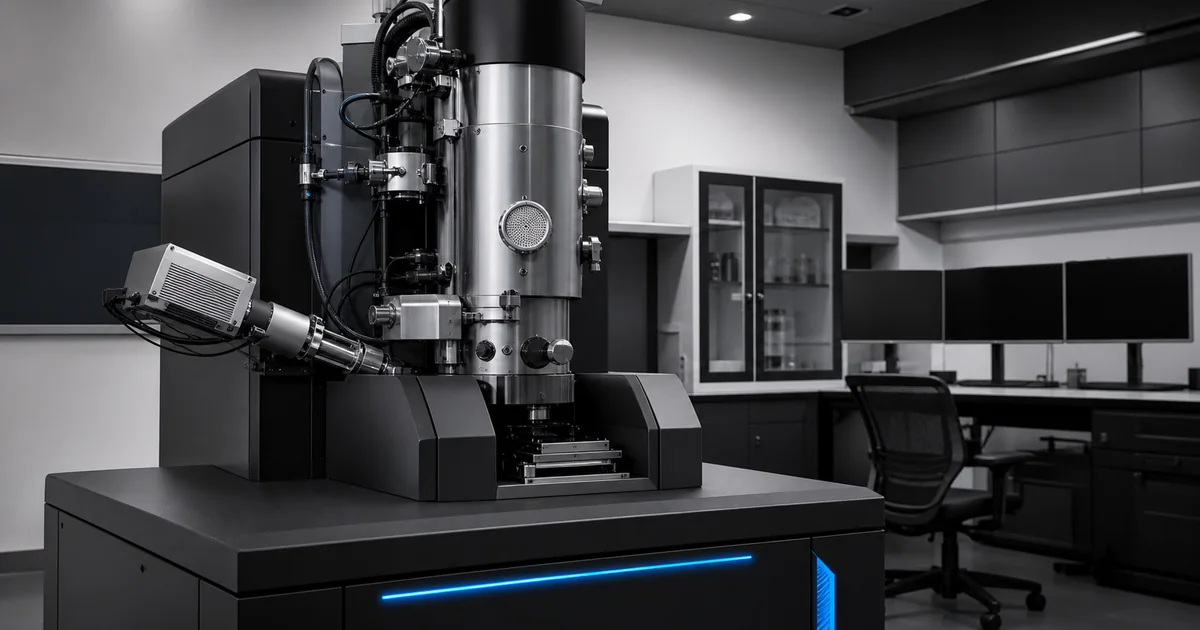
Quick answer
SEM astigmatism is a beam-shape problem that prevents fine detail from becoming sharp in all directions at the same time. A correctly focused but astigmatic image still looks smeared, stretched, or directionally soft.
The fix is stigmation: adjust the X and Y stigmators while focusing on a fine feature until detail is crisp and symmetric.
Key takeaways
- Astigmatism often becomes obvious only at higher magnification.
- A feature that stretches in one direction and then another as focus changes is a classic sign.
- Correct stigmation on a small, sharp, high-contrast feature.
- Refocus after every meaningful stigmator adjustment.
- If the image drifts, charges, or damages quickly, stigmation becomes harder to judge.
Why astigmatism happens
In an ideal SEM, the electron probe is round and small at the sample. Astigmatism makes the probe slightly elongated, so the beam focuses differently in different directions.
Common contributors include:
- aperture contamination
- imperfect aperture alignment
- magnetic or electrostatic asymmetry
- column contamination
- poor beam alignment
- sample charging
- operation at demanding high magnification
Some causes are normal and corrected routinely. Others indicate that the instrument needs cleaning, alignment, or service.
What it looks like
Astigmatism can make particles, pores, scratches, or grain edges look stretched. As you adjust focus through the best point, the direction of blur may rotate. The image never reaches a clean, balanced sharpness.
This is different from simple defocus. Defocus usually makes the whole image soft. Astigmatism makes sharpness directional.
How to correct SEM astigmatism
Use this workflow:
- Move to a stable, detailed feature.
- Increase magnification above the final intended magnification.
- Focus as well as possible.
- Adjust one stigmator axis slowly.
- Refocus.
- Adjust the other axis.
- Refocus again.
- Repeat until fine detail looks equally sharp in all directions.
Do not spin the controls quickly. Small changes are easier to interpret.
Choose the right feature
Good features for stigmation include:
- small particles
- sharp cracks
- coating grains
- pores
- rough fracture edges
- fine surface texture
Poor features include smooth polymers, flat polished areas, low-signal regions, drifting contamination, and charging samples.
Common mistakes
One mistake is correcting astigmatism at low magnification, then assuming it is fine at high magnification. Another is trying to stigmate while the sample is charging. A third is mistaking scan noise for sharpness.
If the image is noisy, slow the scan or average frames before making final judgments.
When correction fails
If stigmation cannot be corrected, check:
- aperture alignment
- aperture cleanliness
- working distance
- beam energy
- sample charging
- stage stability
- contamination buildup
If multiple good samples show the same uncorrectable behavior, the instrument may need column maintenance or service alignment.